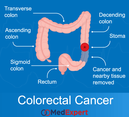
COLORECTAL CANCER
Cancerous tumor growing at the very last part of the gastrointestinal tract is referred to as colorectal cancer. As it is clear from its` name, this type of cancer invades colon and rectum. Colorectal cancer usually starts from a colon polyp, which turns malignant over time. Colorectal cancer symptoms may not be experienced on early stages until the cancer is advanced. Colorectal cancer treatment includes three main options, which are surgery, chemotherapy and radiation. Below we are going to discuss this condition, symptoms is causes, as well as diagnostic procedures and colorectal cancer treatment options, in more details.
Water is absorbed and food residue is converted into waste (faeces) by bacteria when food enters the colon. The part of the colon that stores faeces before it is expelled through the anus is known as the rectum.
Polyps may appear on the inner wall of the colon and rectum. These are benign lumps that are fairly common amongst people aged 50 and above. Certain types of polyps may turn into colorectal cancer and should be removed if they are detected.

Colorectal cancer means that the colon and rectum (or large intestine) which are the last part of the gastrointestinal tract, become affected with cancer cells.
WHAT ARE THE CHARACTERISTICS OF A POLYP THAT MAY INDICATE MALIGNANCY?
- The size of the polyps is greater than 1cm in diameter
- Sessile polyps (no stalks)
- Multiple polyps
Cancer cells are confined to the colon in the early stages of colorectal cancer. The cancer will develop and spread into the lumen of the colon if it is undetected. It will also spread through the colon wall and affect:
- Neighbouring intestines and organs
- The lymphatic system and travel into neighbouring lymph glands (mesenteric lymph nodes)
- The blood stream and travel into the liver where secondary malignant deposits may form

COLORECTAL CANCER SYMPTOMS
Even though colorectal cancer has no symptoms at an early stage, there are warning signs that you should look out for. They include:
- A change in bowel habits
- Blood in stool
- Pain or discomfort in your abdomen
- Anaemia (low red blood cell count)
- Lump in the abdomen
COLORECTAL CANCER TREATMENT OPTIONS
The three treatments mainly used to treat colorectal cancer are surgery, chemotherapy and radiation therapy. These options may be used alone or in a combination.
Surgery
During a colostomy, a new opening (stoma) is created in the abdomen which will then be connected to one end of the colon to allow discharge of faeces. You will be required to wear a colostomy bag to collect faeces and also to learn how to clean and care for the stoma and surrounding skin. This procedure may be temporary or permanent.
Surgery is the main type of treatment for colorectal cancer. The goal of surgery is to completely remove the cancerous sections of the colon and/or rectum as well as the surrounding tissue and mesenteric lymph nodes. After the removal, the two unconnected ends of the bowel are joined (anastomosis). A colostomy diverts the faeces and allows the anastomosis to heal. Once it has healed, the colostomy is closed in a second operation.
Rectal cancers located near the anus may involve complete removal of the anus as well which may require permanent colostomy in the lower right abdomen.
To prevent permanent colostomy, new surgical techniques have been developed to retain the anus. The first technique involved performing intestinal anastomosis with mechanical staples instead of hand sewn stitches (sphincter saving surgery) as close to the anus as possible. The second technique involved creating a new anus using thigh muscles (neo-sphincter surgery).
Surgeries in unique situations include:
- Liver metastases: Removal of liver metastases and excision of the cancerous areas can be performed together if the patient’s overall health allows this. However, this is normally not viable. As a result, patients with liver metastases and colorectal cancer are commonly treated with chemotherapy.
- Large inoperable tumor: If a tumor in the colon or rectum grows too large, intestinal bypass surgery will be required to relieve bowel obstruction.
Lapreoscopic colon surgery (Keyhole surgery)
This minimally invasive technique has been used to treat colorectal cancer. The advantage of this surgery is less pain and faster post-operative recovery. However, it is time consuming, technically difficult and expensive to perform. The risk of cancer implantation in the skin may also be increased. At this stage, this technique is still under clinical evaluation.
Chemotherapy
The administration of toxic drugs to kill cancer cells that can be found at the cancer site or elsewhere in the body after surgery.
It is indicated for patients with advanced colorectal cancer at stages two, three and four after surgery and it also improves survival rates.
Radiotherapy
The administration of powerful radioactive rays to kill cancer cells. This treatment is secondary to surgical excision and is used in these situations:
- After the removal of a colorectal cancer that has affected nearby organs and/or surrounding lymph nodes to kill residual cancer cells within the perimeters of the cancer.
- In the pre-operative phase, it is used on a locally advanced rectal cancer to shrink the tumor so it can be surgically removed.
This treatment is performed in daily sessions, five days a week for four to six weeks. The side effects are normally tolerable and temporary and can range from:
- Abdominal pain and cramps
- Constipation or diarrhoea
- Cystitis
- Excoriation of perianal skin
- Generalized tiredness
DIAGNOSING COLORECTAL CANCER
- History taking and physical examination: Your doctor will ask you questions about your overall health, symptoms you have and family medical history, as those, who have a family history of cancers are assumed to be more predisposed to have the same condition. Then your doctor will perform a physical examination of your body to look for any visible symptoms.
- Colonoscopy: This procedure allows the colonoscopist (doctor who specializes in performing this test) to see inside the entire rectum and colon while you are sedated. If colorectal cancer is detected, a complete diagnosis that can accurately describe the location and stage of cancer may not be possible until the tumor in surgically removed.
- Biopsy: The removal of a small amount of tissue to be examined under a microscope. Even though other tests can suggest the presence of cancer, only a biopsy can make a definite diagnosis. A pathologist (doctor who specializes in lab tests and evaluate cells, tissues and organs to diagnose diseases) then examines the sample. A biopsy may be completed during a colonoscopy or it may be performed on the tissues that have been removed during surgery. A CT scan or ultrasound is sometimes used to guide a needle biopsy. This procedure removes tissue through the skin with a needle that is guided into the tumor.
- Molecular testing of the tumor: Lab tests on the tumor sample may be recommended to detect specific genes, proteins and other factors unique to the tumor. The results of these tests will help to decide if targeted therapy is a treatment option.
- Blood tests: Patients may become anaemic as colorectal cancer often bleeds into the large intestine or rectum. A test of the number of red cells in your blood under the complete blood count (CBC) will be able to indicate whether bleeding is occurring.
Another blood test that reads the levels of a protein called carcinoembryonic antigen (CEA) can indicate if the cancer has spread to other parts of the body. CEA is not the most suitable test for colorectal cancer as levels are high for only about sixty percent of people with the disease that has spread to other organs. Furthermore, other medical conditions can result in an increased CEA. It is often used to monitor colorectal cancer for patients who are already receiving treatment and is not useful as a screening test.
- Computed tomography (CT or CAT) scan: A three dimensional picture of the inside of your body is created using X-rays snapped at different angles. These images are then combined using a computer to produce detailed, cross-sectional view that shows any tumors or abnormalities. It can also be used to measure the size of the tumor. A special dye called contrast medium is sometimes administered before a scan to produce better detail on the image. This dye is injected or given as a pill to swallow. A CT scan can be used on colorectal cancer patients to check if the cancer has spread to the lungs, liver and other organs. This is often conducted in the pre-operative stage.
- Magnetic resonance imaging (MRI): Magnetic fields (not X-rays) are used to produce detailed images of the body. It can also be used to measure the size of the tumor. Contrast medium is also administered before the scan to create a clearer picture. This dye is either injected or given as a pill to swallow. MRI is the best imaging test to locate where the colorectal cancer originated from.
- Ultrasound: Sound waves are used to create an image of the internal organs to determine if the cancer has spread. It can also be used to see the liver, although CT scans are preferred as they better at locating tumors in the liver. Endorectal ultrasound is commonly performed to find out how deep the rectal cancer has developed. It is also used to help plan the treatment. However, this test can’t accurately identify cancer that has spread to nearby lymph nodes or beyond the pelvis.
- Chest x-ray: An image of your internal structures is created using a small dose of radiation. A chest X-ray can help your doctor to determine if the cancer has spread to your lungs.
- Positron emission tomography (PET) or PET-CT scan: A CT scan and a PET scan are normally combined, called a PET-CT scan. However, your doctor may refer to this procedure as a PET scan. This procedure creates images of your organs and tissues inside your body. You will be injected with a small amount of radioactive sugar substance that will be absorbed by cells that use the most energy in your body. Since cancer cells actively use the most energy, it will absorb the radioactive substance. A scanner will then detect this substance to create images of the inside of your body. This procedure is not regularly used for all patients. However, there are specific situations where you doctor may find this procedure useful.


